Home
Uncategories
Female Internal Reproductive Organs Anatomy / Labeled Female Reproductive System Diagram Jpg The Oncofertility Consortium : The female reproductive anatomy includes both external and internal structures.
Female Internal Reproductive Organs Anatomy / Labeled Female Reproductive System Diagram Jpg The Oncofertility Consortium : The female reproductive anatomy includes both external and internal structures.
Female Internal Reproductive Organs Anatomy / Labeled Female Reproductive System Diagram Jpg The Oncofertility Consortium : The female reproductive anatomy includes both external and internal structures.. 3d anatomy tutorial on the female reproductive system from anatomyzone for more videos, 3d models and notes visit: Female organs picture of female reproductive system diagram 1024×1204. Do you know the bones of the skull?. Fetal tissues begin in an undifferentiated state, and based on genetic signals and the interuterine environment the reproductive organs usually differentiate into. That being said, any part of your body can be sexual.
Female reproductive organs undergo substantial structural and functional changes every month. They are present in pairs with the long axis oriented downward and forward. Test your knowledge of the bones of the full skeleton. Can you name the main anatomical areas of the brain?. All anteroposterior diameters are short.
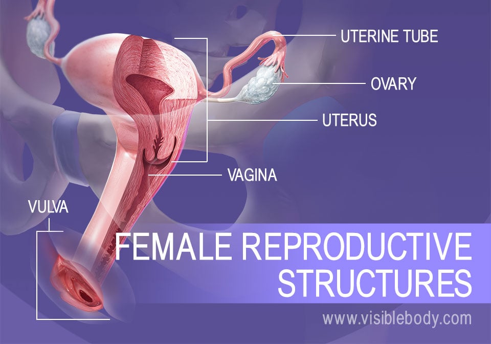
The Female Reproductive System Produces Eggs Facilitates Fertilization And Supports The Developing Embryo from www.visiblebody.com The female reproductive anatomy includes both external and internal structures. Its anatomical structure can be broken down further into the mons pubis. Fetal tissues begin in an undifferentiated state, and based on genetic signals and the interuterine environment the reproductive organs usually differentiate into. All transverse diameters are long. Female organs picture of female reproductive system diagram 1024×1204. During embryonic development the male and female fetus are indistinguishable before about 10 weeks of pregnancy. The major organs of the female reproductive system are located inside the pelvic cavity. The female reproductive tract is all located within the pelvis.
Female reproductive organs undergo substantial structural and functional changes every month.
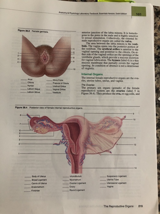
Do you know the bones of the skull?. It is made of muscle and can stretch and grow. The passageway between the outside of the female body and the uterus. The uterus and ovaries are particularly affected by atrophy (shrinkage) after the menopause. In virgins, the hymen usually encircles the opening like a tight ring, but it may completely cover the opening. All anteroposterior diameters are short. The female anatomy consists of female parts, which are external and internal. Posted on june 7, 2016 by admin. The ovaries are the primary organs of the female reproductive system. All transverse diameters are long. It serves important functions during pregnancy and childbirth. Its anatomical structure can be broken down further into the mons pubis. During embryonic development the male and female fetus are indistinguishable before about 10 weeks of pregnancy.
In virgins, the hymen usually encircles the opening like a tight ring, but it may completely cover the opening. During embryonic development the male and female fetus are indistinguishable before about 10 weeks of pregnancy. All transverse diameters are long. Anatomy of female reproductive organs by amrit kaur 2. Name the parts of the human heart
Solved Anatomy Physiology Laboratory Textbook Essentials Chegg Com from media.cheggcdn.com It is made of muscle and can stretch and grow. The 10 most popular quizzes : The function of the external female reproductive structures (the genital) is twofold: Looking at the anatomy of the internal structures of the female reproductive system, i've split this into two parts. The major organs of the female reproductive system are located inside the pelvic cavity. That being said, any part of your body can be sexual. Learn the anatomy of a typical human cell. Fetal tissues begin in an undifferentiated state, and based on genetic signals and the interuterine environment the reproductive organs usually differentiate into.
The major organs of the female reproductive system are located inside the pelvic cavity.
All anteroposterior diameters are short. It is a flat female type. 13.3 commonalities between male and female reproductive anatomy. How about the bones of the axial skeleton?. These organs participate in several hormonal and. Do you know the bones of the skull?. Let's cover the major organs of the pelv. The uterus and ovaries are particularly affected by atrophy (shrinkage) after the menopause. That being said, any part of your body can be sexual. They are present in pairs with the long axis oriented downward and forward. Name the parts of the human heart During embryonic development the male and female fetus are indistinguishable before about 10 weeks of pregnancy. Fetal tissues begin in an undifferentiated state, and based on genetic signals and the interuterine environment the reproductive organs usually differentiate into.
It also is known as the birth canal. These organs are supported in the pelvis by ligaments. All anteroposterior diameters are short. In virgins, the hymen usually encircles the opening like a tight ring, but it may completely cover the opening. These organs participate in several hormonal and.
Female Internal Anatomy Anatomy Drawing Diagram from biology-forums.com Fetal tissues begin in an undifferentiated state, and based on genetic signals and the interuterine environment the reproductive organs usually differentiate into. The vagina, figure 1, is a muscular canal (approximately 10 cm long) that serves as the entrance to the reproductive tract. The passageway between the outside of the female body and the uterus. The female reproductive system is a complex and interconnected group of organs. Test your knowledge of the bones of the full skeleton. It is a flat female type. Its anatomical structure can be broken down further into the mons pubis. Do you know the bones of the skull?.
How about the bones of the axial skeleton?.
It is made up of the vulva, the vagina, the cervix, the uterus, the fallopian tubes and the ovaries. An female's internal reproductive organs are the vagina, uterus, fallopian tubes, cervix, and ovary. Female anatomy includes the external genitals, or the vulva, and the internal reproductive organs, which include the ovaries and the uterus. The major organs of the female reproductive system are located inside the pelvic cavity. Anatomy of female reproductive organs by amrit kaur 2. During embryonic development the male and female fetus are indistinguishable before about 10 weeks of pregnancy. The uterus and ovaries are particularly affected by atrophy (shrinkage) after the menopause. Its anatomical structure can be broken down further into the mons pubis. 13.3 commonalities between male and female reproductive anatomy. Looking at the anatomy of the internal structures of the female reproductive system, i've split this into two parts. Introduction • the reproductive organ in female are those which concerned with copulation, fertilization, growth and development of fetus and its subsequent exit to the outer world. Fetal tissues begin in an undifferentiated state, and based on genetic signals and the interuterine environment the reproductive organs usually differentiate into. The sole aim of this group of organs is to prepare a woman for normal pregnancy and its maintenance till the child is delivered.
The 10 most popular quizzes : female internal. The female reproductive tract is all located within the pelvis.

Innovative information,
BalasHapusSexologist in Allahabad